Home
/ Compact Bone Diagram Microscope - The Skeletal System Cls 224 Essentials Of Human - This video describes the microscopic anatomy of compact bone.
Compact Bone Diagram Microscope - The Skeletal System Cls 224 Essentials Of Human - This video describes the microscopic anatomy of compact bone.
Compact Bone Diagram Microscope - The Skeletal System Cls 224 Essentials Of Human - This video describes the microscopic anatomy of compact bone.. Online quiz to learn compact bone microscope slide labeled ; These bones tend to support weight and. The compact bone is the main structure in the body for support, protection, and movement. Due to the strong nature of compact bone, compared to spongy bone, it is the preferred tissue for strength. Do you want to learn the details of the histology of compact bone with labelled diagram and authentic slide images?
The spaces between the trabeculae contain red or yellow marrow, depending on a person's age and on which bone it is. A diagram of the anatomy of a bone, showing the compact bone. Compact bone is formed in concentric circles. Transverse section of an osteon with its haversian canal [1. Under the microscope, bone can be divided into two types compact bone forms the outer 'shell' of bone.
Cartilage Bone Ossification The Histology Guide from www.histology.leeds.ac.uk Spongy bone is used for more active functions of the bones, including blood cell production and ion exchange. To prepare this slide, a bone specimen is ground thin and then. The central canal, lamellae, canaliculi, and lacunae with osteocytes are apparent. Microscopic structure of a long bone. Like other tissues in the body, bones are made up of specialized cells that serve different functions. Compact and spongy bone with dr. You can think of compact bone as being very similar. Before placing your slide on the microscope stage, remember to read the label, examine the slide with your eye and note any visible macroscopic features that might help your examination.
The remainder is cancellous bone, which has a spongelike appearance with numerous large spaces and is found in the.
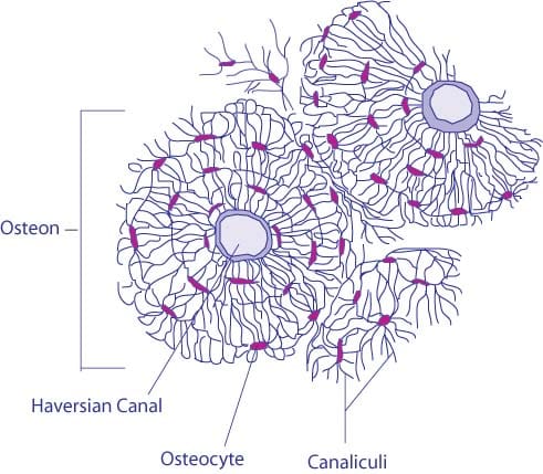
Start studying compact bone under microscope. The larger ovals are blood vessels running through the bone. Although the calls are close together, this type of bone is not completely solid. 0 0000 a shoutout is a way of letting people know of a. At this level of magnification, the fundamental structure of compact bone is visible. Like other tissues in the body, bones are made up of specialized cells that serve different functions. Before placing your slide on the microscope stage, remember to read the label, examine the slide with your eye and note any visible macroscopic features that might help your examination. Start studying compact bone microscopic labeling. The spaces between the trabeculae contain red or yellow marrow, depending on a person's age and on which bone it is. Bone tissue and cells under the microscope introduction. The central canal, lamellae, canaliculi, and lacunae with osteocytes are apparent. This human bone section shows the haversian canal (or osteon) structure of compact bone tissue. To prepare this slide, a bone specimen is ground thin and then.
Good, here in this part, i am going to describe the structure of compact bone. Each osteon looks like a ring with a light spot in the center. This type of bone is located between layers of compact bone and is thin and porous. This video describes the microscopic anatomy of compact bone. 0 0000 a shoutout is a way of letting people know of a.
What Is The Microscopic Anatomy Of A Compact Bone Quora from qph.fs.quoracdn.net Under magnification you can clearly see the system of concentric circles that forms compact bone. It is enveloped by lamellae in the ground substance, which may be more or less impregnated with silver nitrate. The larger ovals are blood vessels running through the bone. Some, mostly older, compact bone is remodelled to form these haversian systems (or osteons). Learn vocabulary, terms, and more with flashcards, games, and other study tools. However, compact bones also serve a function in storing and releasing calcium to the. You can think of compact bone as being very similar. The light spot is a canal that carries a blood vessel and a nerve fiber.
The collagen fibers in the more heavily stained lamellae are arranged in a circular fashion;
Compact bone is formed in concentric circles. The spaces between the trabeculae contain red or yellow marrow, depending on a person's age and on which bone it is. 100x on this image you can see several of the structural units of bone tissue (osteons or haversian systems). Compact bone, also called cortical bone, dense bone in which the bony matrix is solidly filled with organic ground substance and inorganic salts, leaving only tiny spaces (lacunae) that contain the osteocytes, or bone cells.compact bone makes up 80 percent of the human skeleton; Compact and spongy bone with dr. Compact bone, or cortical bone, mainly serves a mechanical function. The cavity that extends the length of the diaphysis is the medullary cavity. The scanning electron microscope (sem) is among the most frequently used instruments for examining bone. (b) in this micrograph of the osteon, you can clearly see the concentric lamellae and central canals. Spongy bone is used for more active functions of the bones, including blood cell production and ion exchange. Students can easily learn the structure of dry, compact bone using this prepared microscope slide. Bone tissue and cells under the microscope introduction. Learn vocabulary, terms, and more with flashcards, games, and other study tools.
Learn vocabulary, terms, and more with flashcards, games, and other study tools. A photo taken through a microscope that shows the anatomy of compact bone with a detailed view of an osteon. Online quiz to learn compact bone microscope slide labeled ; The scanning electron microscope (sem) is among the most frequently used instruments for examining bone. Good, here in this part, i am going to describe the structure of compact bone.
Ultrastructure Of Bone Components Structure Teachmeanatomy from teachmeanatomy.info Compact bone, or cortical bone, mainly serves a mechanical function. Online quiz to learn compact bone microscope slide labeled ; The central canal, lamellae, canaliculi, and lacunae with osteocytes are apparent. The spaces between the trabeculae contain red or yellow marrow, depending on a person's age and on which bone it is. The cells of compact bone, which is also called cortical bone, appear to be tightly packed into a solid mass. The darker ring consists of layers of bone matrix made by cells called. Transverse section of an osteon with its haversian canal [1. The scanning electron microscope (sem) is among the most frequently used instruments for examining bone.
Compact bone, also called cortical bone, dense bone in which the bony matrix is solidly filled with organic ground substance and inorganic salts, leaving only tiny spaces (lacunae) that contain the osteocytes, or bone cells.compact bone makes up 80 percent of the human skeleton;
100x on this image you can see several of the structural units of bone tissue (osteons or haversian systems). Like other tissues in the body, bones are made up of specialized cells that serve different functions. Compact bone, or cortical bone, mainly serves a mechanical function. Do you want to learn the details of the histology of compact bone with labelled diagram and authentic slide images? Bone tissue and cells under the microscope introduction. Under the microscope, bone can be divided into two types compact bone forms the outer 'shell' of bone. 0 0000 a shoutout is a way of letting people know of a. However, compact bones also serve a function in storing and releasing calcium to the. The scanning electron microscope (sem) is among the most frequently used instruments for examining bone. Students can easily learn the structure of dry, compact bone using this prepared microscope slide. The compact bone is the main structure in the body for support, protection, and movement. Under magnification you can clearly see the system of concentric circles that forms compact bone. If you look at compact bone under the microscope, you will observe a highly organized arrangement of concentric circles that look like tree trunks.
The compact bone is composed of calcified extracellular material the bone matrix and 3 major cell types which are osteoblast which ssynthesize and secrete the organic components of bone matrix which include type 1 collagen fibers proteoglycans and several glycoproteins such as ostepnectin compact bone diagram. You can think of compact bone as being very similar.
